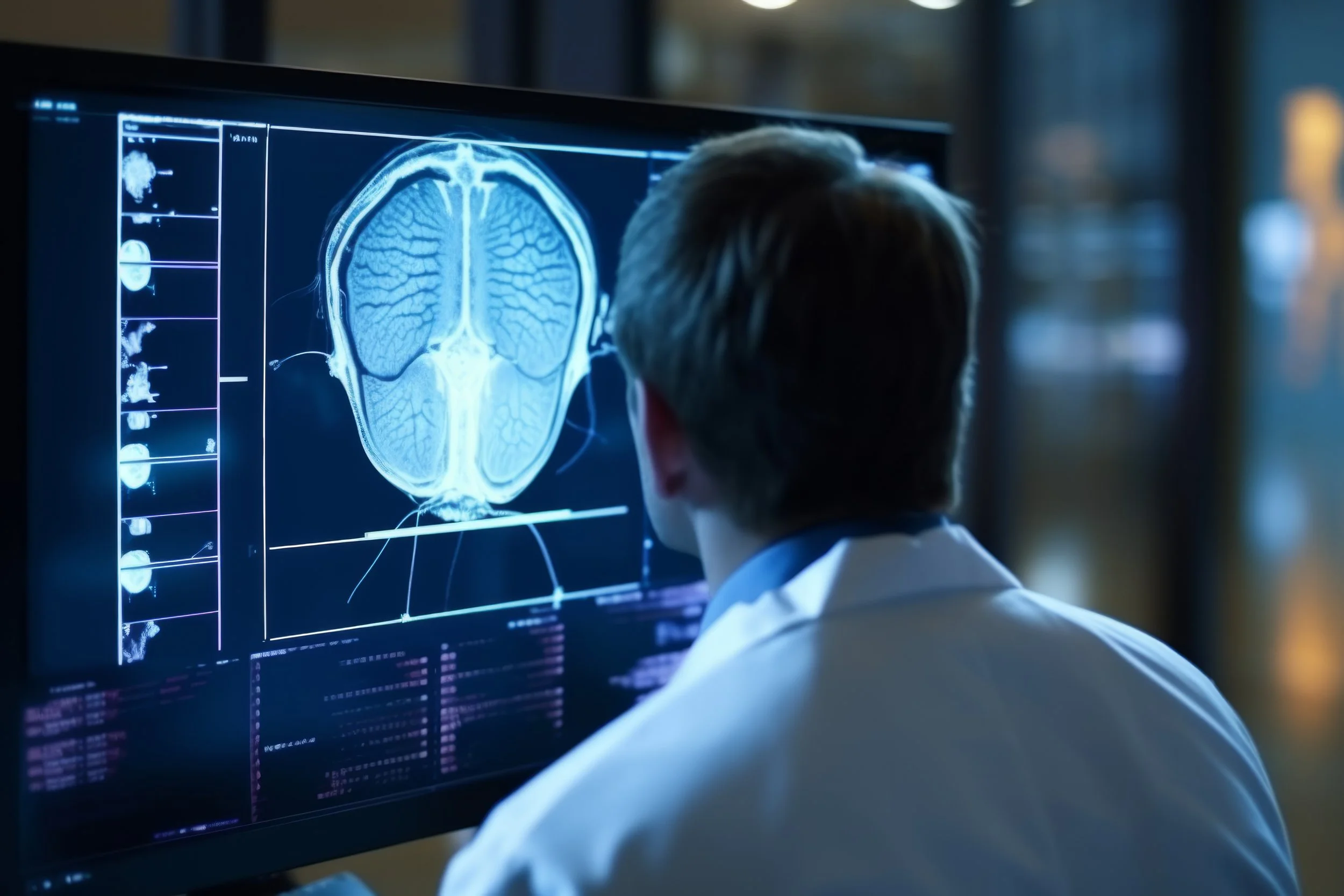
3.0T MAGNETIC RESONANCE IMAGING (MRI)
MRI (Magnetic Resonance Imaging) utilizes a strong magnet, radio waves, and a computer to create detailed, cross-sectional images of internal organs and structures. MRI is particularly useful for capturing detailed images of internal organs and tissues, blood vessels, and bone structure. These images provide crucial information to your doctor that helps him or her make accurate diagnoses and treatment plans for you. Depending on the exams, intravenous contrast may be given under certain indications to help improve the visualization of certain structures.
3.0T MRI is extremely efficient for the fastest, highest-quality scans and a greater range of imaging capabilities. Utilizing shorter scan times, the 3T machine maximizes patient comfort without compromising quality. The superb reliability of high-field MRI allows our board-certified radiologists to differentiate between benign and potentially hazardous medical conditions with confidence. This allows your healthcare team to provide you with earlier diagnosis and treatment, subsequently leading to more positive outcomes.
WHAT ARE SOME COMMON USES OF 3.0T MRI?
The development of the MRI scan represents a huge milestone for the medical world.
Doctors, scientists, and researchers are now able to examine the inside of the human body in high detail using a non-invasive tool.
The following are examples in which an MRI scanner would be used:
anomalies of the brain and spinal cord
tumors, cysts, and other anomalies in various parts of the body
breast cancer screening for women who face a high risk of breast cancer
injuries or abnormalities of the joints, such as the back and knee
certain types of heart problems
diseases of the liver and other abdominal organs
the evaluation of pelvic pain in women, with causes including fibroids and endometriosis
suspected uterine anomalies in women undergoing evaluation for infertility
HOW SHOULD I PREPARE for this exam?
Before your MRI exam, remove all accessories, including hair pins, jewelry, eyeglasses, hearing aids, wigs, and dentures. During the exam, these metal objects may interfere with the magnetic field, affecting the quality of the MRI images taken. Notify your technologist if you:
have any prosthetic joints – hip, knee
have a heart pacemaker (or artificial heart valve), defibrillator, or artificial heart valve
have an intrauterine device (IUD)
have any metal plates, pins, screws, or surgical staples in your body
have neuro-stimulators
have inner ear implants
have tattoos and permanent make-up
have a bullet or shrapnel in your body, or ever worked with metal
are pregnant or suspect you may be pregnant
WHAT can I EXPECT DURING THIS PROCEDURE
Once in the scanner, the MRI technician communicates with the patient via the intercom to ensure comfort. They will not start the scan until the patient is ready.
During the scan, it is vital to stay still. Any movement will disrupt the images, much like a camera trying to take a picture of a moving object. The scanner will make loud noises; this is perfectly normal. Depending on the images, at times, it may be necessary for the person to hold their breath.
If the patient feels uncomfortable during the procedure, they can speak to the MRI technician via the intercom and request that the scan be stopped.
WHAT ARE THE BENEFITS OF 3.0T MRI?
A more accurate diagnosis leading to better outcomes
Shorter examination times; a patient with claustrophobia can receive a brain MRI in as little as fifteen minutes with the 3.0T MRI, reducing the time you spend in an MRI machine, reducing your stress levels
More detailed images with greater resolution enable the identification of smaller lesions and anatomical structures
Detects problems that cannot be seen with less powerful scanners
Allows more sophisticated imaging procedures
Has a lower risk of distorted images, so it lessens the need for repeated scans
Reduces the need for invasive procedures


