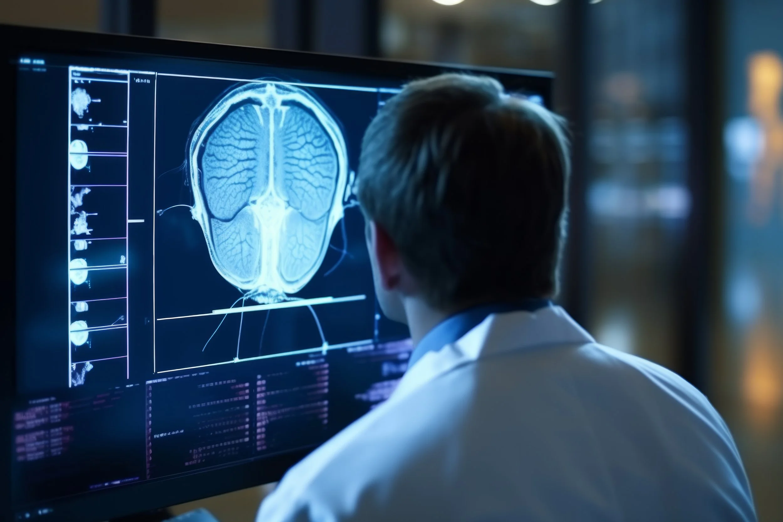
NUclear Medicine
Nuclear medicine involves the use of very small amounts of radiopharmaceuticals to diagnose disease. The procedures are safe, involve little or no patient discomfort, and do not require the use of anesthesia.
Rezolut utilizes the most advanced nuclear imaging tools available to help providers safely and effectively diagnose and treat a variety of conditions.
HOW NUCLEAR MEDICINE WORKS
Nuclear medicine imaging involves tracing very small amounts of radioactive materials in the bloodstream. These materials are injected, swallowed, or inhaled, depending on the area needing examination. These “radiotracers” give off gamma rays and are absorbed by specific bones, organs, and tissues. The technologist uses a special camera and a computer to track the gamma rays and create images of internal body structures, function, and physiology. This provides highly unique information that a provider would be unable to obtain using other imaging options and makes it possible to detect diseases at their very earliest stage.
The radioactive compounds have few to no side effects, and the tests are completely painless, safe, and noninvasive.
APPLICATIONS FOR NUCLEAR MEDICINE
Nuclear medicine is used primarily to diagnose diseases and disorders and to assess body or organ function. It has been used in all major systems of the body, including the heart, lungs, bones, and brain, to assess:
Blood Clots
Blood Flow and Function
Cancer Treatment Response
Cardiac Function and Health
Coronary Artery Disease
Liver and Gallbladder Function
Lung Function
Neurological Abnormalities
Orthopedic Injuries and Infections
Thyroid Function and Disease
Transplant Rejection
Unexplained Bleeding
WHAT CAN I EXPECT DURING THIS PROCEDURE?
Although imaging time can vary, the exam generally takes 20 to 45 minutes. A radiopharmaceutical, known as a tracer, is usually administered either intravenously or by mouth. What radiopharmaceutical is used and when is dependent upon the type of exam you’re having. For most nuclear scans, you will lie down on a table, and a nuclear imaging camera will be used to capture the image of the area being examined. The camera is either suspended over or below the exam table or in a large donut-shaped machine similar to a CT scanner. While the images are being obtained, you must remain as still as possible. Most of the radioactivity is expelled out of your body in urine or stool. The rest simply disappears over time.
WHAT WILL I EXPERIENCE DURING THE PROCEDURE?
Although intravenous injections are usually done with a small needle, some patients experience minor discomfort from the injection. Also, lying still on the examining table may be uncomfortable for some patients. You will hear low-level clicking or buzzing noises from the machine.


