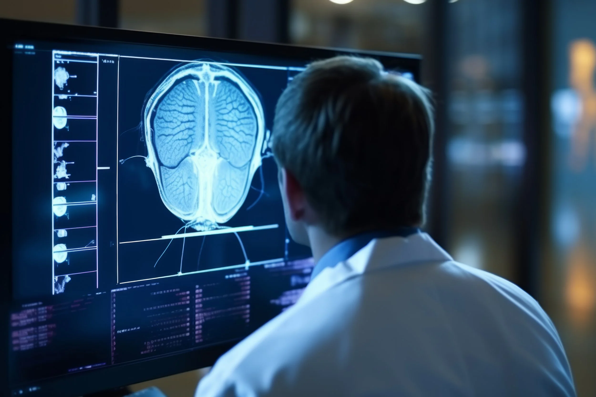
PET/CT
Positron emission tomography (PET) is a nuclear medicine exam that produces a 3D image of functional processes in the body. A PET scan uses a small amount of a radioactive drug to show differences between healthy and diseased issues. The diagnostic images produced by PET are used to evaluate a variety of diseases. PET and CT are used to exam internal organs, tissues, and bones, but do so in different ways. A PET scan measures quantifiable changes in metabolic activity, including blood flow and oxygen use. A CT scan, by contrast, functions more like an advanced X-ray. Multiple images are taken in rapid succession and from different angles to produce a cross-sectional view of the area in question.
WHEN PET/CT SCANS ARE USED
PET/CT scanning is quickly becoming the ideal choice for diagnosing many cancers and cardiac and neurological abnormalities. The scan can not only help diagnose conditions but is also instrumental in helping physicians track the effectiveness of treatment. Applications for PET/CT include:
Brain Changes and Conditions
Blood Distribution and Flow, including Blood Clots and Strokes
Cardiac Conditions, Health, and Performance
Cancer Diagnosis and Tracking
Neurological Disorders, Including Seizures and Dementia
Tissue and Tumor Diagnosis
BENEFITS OF PET/CT SCANNING
The combined scan provides physicians with two crucial pieces of information: changes in cellular function and the location of abnormalities, providing greater diagnostic accuracy than either scan alone. This information helps physicians determine the best course of treatment by pinpointing the location of the disease and its effects on the body.
HOW SHOULD I PREPARE FOR THIS PROCEDURE?
PET is usually done on an outpatient basis. You should wear comfortable clothes. You will be instructed not to eat for four to six hours before your scan. Drink plenty of water prior to your exam. Consult with your doctor regarding the use of medications before the test.
WHAT SHOULD I EXPECT FROM THIS PROCEDURE?
You will receive an intravenous (IV) injection of the radioactive substance. The radioactive substance will then take approximately 30 to 90 minutes to travel through your body and be absorbed by the tissue under study. During this time, you will be asked to rest quietly and avoid significant movement or talking, which may alter the localization of the administered substance. You will be positioned on the PET scanner table and be asked to lie still during your exam. Scanning takes 30 to 45 minutes.
Some patients being evaluated for heart disease may undergo a stress test in which PET scans are obtained while they are at rest and again after a pharmaceutical is administered to alter the blood flow to the heart. Usually, there are no restrictions on daily routine after the test. You should drink plenty of fluids to flush the radioactive substance from your body.
WHAT WILL I EXPERIENCE DURING THE PROCEDURE?
If given an intravenous injection, you will feel like a slight prick. However, you will not feel the substance in your body. You will be made as comfortable as possible on the exam table before you are positioned in the PET scanner for the test. You will hear buzzing or clicking sounds during the exam. Claustrophobic patients may feel some anxiety while positioned in the scanner. Some patients find it uncomfortable to hold still in one position for more than a few minutes.


